

Santosh Manicka and Michael Levin Introduce Topoiesis in Collective Morphogenesis
How can a group of developing embryos survive a chemical insult that kills every isolated individual, without relying on a central pacemaker or specialized organizer cells? In their November 3, 2026 preprint A topoietic model of collective morphogenesis and compartmentalization (bioRxiv 2026.11.03.763900, doi:10.64898/2026.11.03.763900), Allen Discovery Center researchers Santosh Manicka and Michael Levin at Tufts University coin the term topoiesis (from the Greek topos for place or space and poiesis for making) to model Cross-Embryo Morphogenetic Assistance (CEMA). Their calibrated biophysical framework shows that macroscale group geometry and extracellular diffusion alone can organize a biological collective, driving embryo survival from 0 percent in solitary Xenopus laevis embryos up to 92 percent in collectives of $N = 306$.
Within the broader investigation into how scientists define and measure consciousness across biological and artificial substrates, Manicka and Levin’s topoiesis model extends the Tufts bioelectric research programme beyond single-body regeneration. While Léo Pio-Lopez and Michael Levin’s September 2026 work on scale-free niche construction and bioelectric stigmergy examined how cells write persistent cues into their local extracellular matrix, topoiesis demonstrates how the physical perimeter of a multi-organism group acts as an organizing sink that couples group size directly to internal bioelectric error correction.
Cross-Embryo Morphogenetic Assistance (CEMA) and the Topoietic Mechanism
In wetlab experiments on Xenopus laevis tadpole development, embryos exposed to teratogenic stress exhibit severe craniofacial defects and complete mortality when raised in isolation ($N = 1$). When identical stressed embryos are placed together in larger dishes, their survival rate rises monotonically with group size, reaching 92 percent at $N = 306$ while craniofacial defect rates drop in parallel.
Classical developmental biology explains spatial patterning through either Wolpert-style positional information gradients emitted by a localized source or Alan Turing’s reaction-diffusion instabilities between an activator and a faster-diffusing inhibitor. Neither mechanism explains how a loose clustering of separate embryos generates a coherent rescue field without an internal organizer.
Manicka and Levin show that topoiesis requires no specialized organizer cell and no two-morphogen Turing instability. Instead, three coupled physical components drive the collective rescue.
- Every stressed embryo constitutively secretes a fast-diffusing extracellular stress cue, modeled on purinergic signaling molecules such as extracellular ATP or adenosine, which diffuses across the shared medium and undergoes first-order decay over a characteristic length scale $\lambda$.
- The outer boundary of the embryo collective acts as an absorbing sink where the diffusive signal escapes into the surrounding bath, while the interior volume retains the secreted molecules. Because interior volume scales faster than perimeter surface area as group radius $L$ grows, the dimensionless ratio $L/\lambda$ governs the steady-state concentration profile across the collective.
- Inside each embryo, a tissue-level bioelectric lattice of cells coupled by voltage-gated gap junctions reads the local extracellular signal concentration. When the collective exceeds the critical $L/\lambda$ threshold, the elevated extracellular field shifts the voltage-gated gap junction conductances into a homeostatic attractor that preserves normal craniofacial pre-patterns against teratogenic perturbation.
| Patterning Framework | Primary Organizing Driver | Boundary Role | Scaling Control Parameter | Biological Example |
|---|---|---|---|---|
| Wolpert Positional Gradient | Localized source secreting a morphogen | Passive tissue edge | Distance from localized organizer | Drosophila Bicoid anterior-posterior axis |
| Turing Reaction-Diffusion | Short-range activator + long-range inhibitor | Reflective or periodic boundary | Diffusivity ratio $D_{\text{inh}} / D_{\text{act}}$ | Vertebrate skin pigmentation stripes |
| Bioelectric Stigmergy (Pio-Lopez & Levin 2026) | Mobile agents depositing extracellular vesicles | Dynamic niche trail | Agent density and trail decay rate | Xenopus and planarian tissue remodeling |
| Topoiesis (Manicka & Levin 2026) | Uniform constitutive secretion + absorbing sink | Active organizing sink defining compartment identity | Group size to decay length ratio $L/\lambda$ | Cross-Embryo Morphogenetic Assistance ($N = 1 \to 306$, $0\% \to 92\%$ survival) |
Compartmentalization and Multiscale Individuation
Beyond explaining why grouped embryos survive, Manicka and Levin test how a continuous field of cells partitions itself into distinct functional compartments. By varying the geometry of the dish boundary and the diffusion decay length $\lambda$, the topoietic model produces discrete interior zones with sharp bioelectric phase boundaries.
This mechanism connects directly to Michael Levin’s cognitive light cone framework and planarian bioelectric memory. In planarian regeneration, temporarily blocking gap junctions with octanol causes a single flatworm bisected in water to regenerate two heads, permanently rewriting the anatomical target memory stored in its bioelectric circuit without altering its genome. In the topoietic model of CEMA, the intervention happens one level higher in the spatial hierarchy. Rather than editing individual gap junctions or individual genes, changing the macroscale geometry of the collective ($L/\lambda$) controls whether the internal bioelectric lattices inside hundreds of separate embryos lock into their healthy target morphology or fragment into teratogenic defects.
What Topoiesis Tells Us About Emerging Consciousness and Self-Boundaries
Why does a biophysical model of frog embryo survival bear on the science of consciousness? Under functionalist emergentism and Enactivism (Varela, Thompson, and Noë), as well as Karl Friston’s Markov blanket formalism, any conscious or proto-cognitive system must first establish a self-boundary that separates internal states from external states.
Standard neurocentric theories treat that boundary as anatomically fixed by the skull or the outer cell membrane. Manicka and Levin’s topoiesis model demonstrates that functional individuation is dynamically negotiated by geometry and field diffusion. When $N = 306$ embryos share an extracellular purinergic field whose gradient is shaped by the collective perimeter, the group acts as a temporary higher-order homeostatic unit whose macroscale boundary stabilizes the microscale bioelectric states of its members.
This finding sharpens the contrast with Ricard Solé and Michael Levin’s October 2026 analysis of large language models as cognitive viruses and Michael Levin’s cognitive light cone model of unconventional substrates. Current disembodied language models have no metabolic or morphogenetic boundary to defend, whereas even pre-neural embryonic collectives construct a shared spatial envelope that performs error correction across hundreds of individuals. Topoiesis does not claim that an embryo dish possesses subjective phenomenal awareness. Instead, it identifies the exact physical threshold where isolated living subunits begin computing as a unified collective self-model.
Comparison to The Consciousness AI
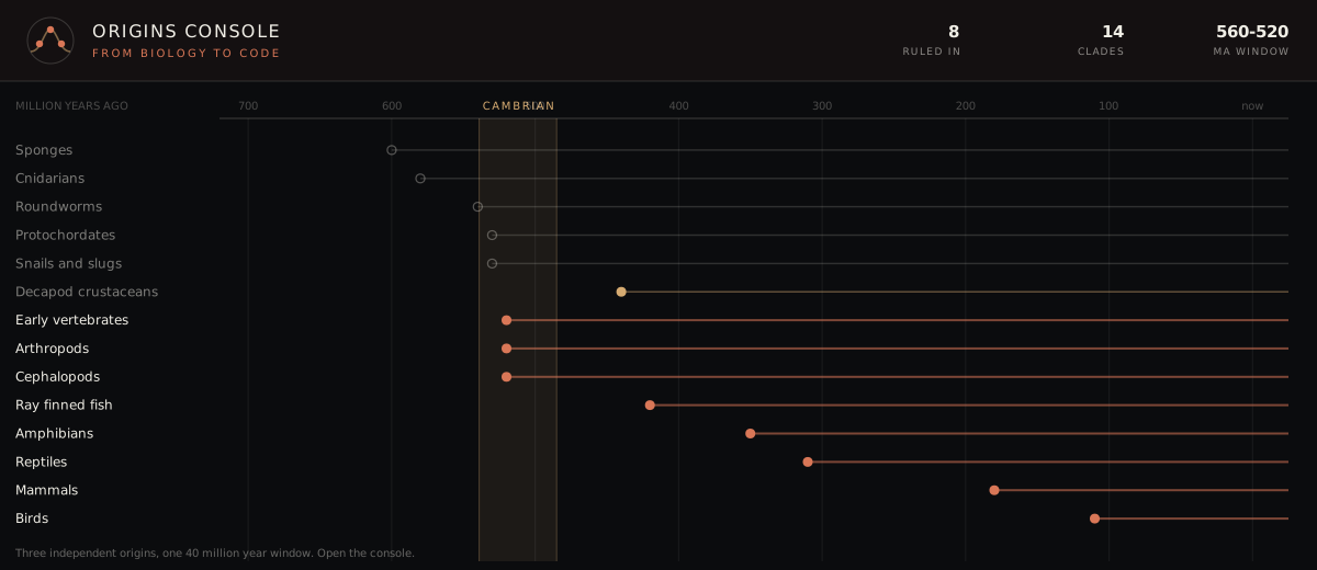
In The Consciousness AI, our Origins Console maps the evolutionary transitions from pre-neural bioelectric coordination and homeostatic boundary maintenance to centralized nervous systems across the Feinberg and Mallatt clades. In our active research codebase, the Existence Guarantee (AE-1) indicator tracks whether an agent actively defends its homeostatic integrity against environmental perturbations rather than following fixed rewards.
As documented in our November 2026 research inventory, AE-1 currently holds a PARTIAL status while competing-goals and drive-off ablation gates are evaluated, and 0 of 21 scalar instruments are marked trusted. Manicka and Levin’s topoietic model suggests a concrete architectural principle for multi-agent and modular neural substrates. Rather than wiring all error-correction signals through a dense central controller, a distributed substrate can regulate local module excitability through a shared, decaying diffusive field whose equilibrium is governed by the active perimeter of the workspace.
Scope Limits and Open Empirical Questions
Two empirical questions remain open in the bioRxiv preprint. First, while the computational lattice reproduces the $N = 1$ to $306$ survival curve and craniofacial defect rates of Xenopus CEMA, identifying the exact molecular ratios between extracellular ATP, adenosine, and downstream ion-channel phosphorylation requires targeted pharmacological knockdowns across varying dish geometries. Second, topoietic coordination relies on slow diffusive transport across millimeter-to-centimeter scales, which suffices for embryonic morphogenesis over hours but must hand off to fast axonal and thalamic broadcasting once an organism develops real-time sensorimotor consciousness.
Researchers covered here
- Santosh ManickaAllen Discovery Center, Tufts UniversityTopoietic models of collective morphogenesis, Cross-Embryo Morphogenetic Assistance, multiscale bioelectric network modeling
-
 Michael LevinAllen Discovery Center, Tufts UniversityBioelectric signalling, the cognitive light cone, and diverse intelligence