Michael Levin and Developmental Bioelectricity Across Non-Neural Tissues From Seconds to Hours
How fast do non-neural living tissues actually compute with voltage, and do their bioelectric patterns merely mirror calcium waves or operate as an independent multi-timescale medium? In a peer-reviewed study published in Developmental Biology (Volume 539, August 2026, doi:10.1016/j.ydbio.2026.08.001), Patrick McMillen and Michael Levin (Allen Discovery Center, Tufts University) apply quantitative Fluorescent Lifetime Imaging (FLIM) to map developmental bioelectricity continuously for more than 17 hours across spreading Xenopus laevis neural crest explants. Their recordings reveal three nested bioelectric timescales running across seconds, minutes, and hours, spanning multiple cells at long range, with minutes-scale voltage dynamics mathematically distinct from calcium signaling.
Within the broader investigation tracked in how scientists race to define and test AI consciousness in 2026 and our Bioelectricity hub, McMillen and Levin’s paper addresses a major empirical gap. Standard neuroscience treats fast millisecond action potentials in neurons as the sole carrier of biological information processing, while viewing non-neural resting potentials as static background gradients. Capturing continuous multi-scale resting potential dynamics over 17 hours shows that somatic tissues run rich, multi-layered voltage computations long before or outside a centralized brain.
Quantitative FLIM Mapping Across 17 Hours of Living Tissue
Traditional intensity-based voltage-sensitive dyes struggle over long developmental windows because photobleaching, dye uptake differences, and tissue movement distort raw brightness readings. McMillen and Levin overcome those artifacts by using Fluorescent Lifetime Imaging (FLIM) to produce an optical estimate of resting membrane potential (V_mem) independent of local dye concentration.
By tracking live Xenopus laevis neural crest explants continuously for over 17 hours, the team captured developmental bioelectricity across three distinct temporal bands operating simultaneously inside the same migrating cell collective.
- A slow hours-scale bioelectric component that tracks long-range tissue-level patterning and anatomical state maintenance.
- An intermediate minutes-scale dynamic component propagating across multicellular clusters.
- A faster seconds-scale component revealing rapid non-neural voltage fluctuations previously blurred out by infrequent time-lapse snapshots.
Crucially, information-theoretic comparison between simultaneous membrane-potential and calcium recordings demonstrated that the minutes-scale V_mem signals are largely distinct from calcium signaling dynamics. Developmental bioelectricity is not a passive readout of calcium waves. Surveying these long-distance events across spreading explants also points toward coordinated cell-to-cell transmission mechanisms that likely include physical coupling via tunneling nanotubes alongside gap junctions.
| Biological Signaling Medium | Characteristic Timescale | Primary Physical Carrier | Role in Collective Tissue Computation |
|---|---|---|---|
| Neuronal Action Potentials | Milliseconds (1 to 10 ms) |
Voltage-gated sodium and potassium spikes along axons | Rapid sensorimotor control and fast cortical binding |
Seconds-Scale Non-Neural V_mem |
Seconds (McMillen & Levin 2026) | Rapid local ion-channel fluctuations measured by FLIM | Short-range excitability and immediate multicellular coordination |
Minutes-Scale Non-Neural V_mem |
Minutes (distinct from calcium) | Propagating membrane-potential shifts across explants | Intermediate collective coordination independent of calcium waves |
| Hours-Scale Developmental Bioelectricity | Hours (>17 h continuous FLIM) |
Stable multicellular V_mem domains and gap junctions |
Long-range anatomical target memory and morphogenesis |
Connecting Lifetime Imaging to Topoiesis, Stigmergy, and Planarian Memory
Reading McMillen and Levin’s Developmental Biology study alongside three recent Tufts University papers covered on this site reveals how the experimental pieces of Levin’s bioelectric research programme fit together.
In Michael Levin’s cognitive light cone model of unconventional substrates and his planarian bioelectric memory and xenobot experiments, altering gap-junction coupling rewrites the anatomical target morphology stored by a flatworm’s tissue without changing its DNA. More recently, Léo Pio-Lopez and Michael Levin’s work on bioelectric stigmergy analyzed how cells write persistent cues into their extracellular niche, and Santosh Manicka and Michael Levin’s November 2026 topoiesis preprint modeled how macroscale dish geometry stabilizes internal embryonic bioelectric lattices across collectives of up to N = 306 embryos.
McMillen and Levin’s 17-hour FLIM recordings supply the missing empirical microscope underneath those computational models. Rather than observing a single flat resting potential that flips once per embryonic stage, live neural crest explants exhibit stacked seconds, minutes, and hours oscillations spanning cell groups via gap junctions and likely tunneling nanotubes.
What Multi-Scale Developmental Bioelectricity Tells Us About Consciousness
Why do non-neural voltage waves in frog neural crest cells bear directly on the science of consciousness? First, they challenge the binary boundary between “non-cognitive somatic chemistry” and “cognitive neural circuits” mapped on our Origins of Consciousness Console. Evolutionary neuroscience already knows that the ion channels, pumps, and electrical synapses powering vertebrate brains originated in pre-neural tissues that solved anatomical navigation problems across hours rather than milliseconds.
Second, the finding tests substrate independence from a biological direction. If basal goal-directed problem solving and collective memory operate over seconds to hours in non-neural cells via ionic V_mem fields, then cognitive integration is neither tied exclusively to millisecond action potentials nor restricted to synaptic wiring alone. McMillen and Levin do not claim that neural crest explants possess subjective phenomenal awareness. Instead, their recordings show how living matter stacks fast, medium, and slow physical timescales in one continuous electrical substrate, illuminating the pre-neural foundation from which vertebrate nervous systems and conscious experience evolved.
Comparison to The Consciousness AI and bioelectrics
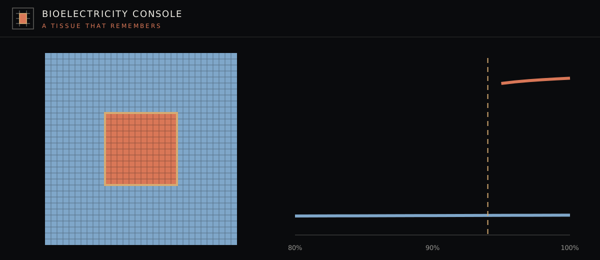
McMillen and Levin’s call for quantitative models that link multi-scale bioelectric parameters directly to tissue-level outcomes aligns closely with our open-source software repository tlcdv/bioelectrics (Zenodo DOI 10.5281/zenodo.23531183). Built in PyTorch and grounded in published somatic models from Michael Levin’s lab, Alexis Pietak (BETSE), and Javier Cervera and Salvador Mafe, bioelectrics simulates non-neural tissue V_mem dynamics at the level of ions, channels, pumps, and voltage-gated gap junctions, while computing exact gradients to solve the bioelectric inverse problem (finding short drug or light interventions that move a tissue into a persistent target V_mem attractor).
We keep strict facts discipline around that connection. bioelectrics is a differentiable software simulation engine whose validation and limitations are published openly in its repository documentation; it does not reproduce wet-lab FLIM measurements on Xenopus neural crest explants and does not yet model tunneling-nanotube transport. Readers can experiment directly with bistable tissue domains and gap-junction coupling computed by the bioelectrics engine inside the interactive console below.
Open Questions for Bioelectric Neuroscience
Two technical frontiers open directly from McMillen and Levin’s Developmental Biology paper. First, proving the exact contribution of tunneling nanotubes versus classical gap junctions in propagating minutes-scale and seconds-scale voltage events will require targeted pharmacological and optogenetic perturbations under continuous FLIM imaging. Second, integrating these newly observed seconds-scale and minutes-scale fluctuations into differentiable tissue models will clarify whether fast non-neural voltage bursts serve as error-correction pulses that steer slower hours-scale morphogenetic attractors, a multi-timescale hierarchy with direct parallels to fast spiking and slow neuromodulation inside conscious brains.
Sources
- Patrick McMillen and Michael Levin. Lifetime imaging reveals long-distance non-neural bioelectric patterns on timescales from seconds to hours. Developmental Biology Volume 539, August 2026. doi:10.1016/j.ydbio.2026.08.001.
- Santosh Manicka and Michael Levin. A topoietic model of collective morphogenesis and compartmentalization. bioRxiv preprint
2026.11.03.763900, November 3, 2026. doi:10.64898/2026.11.03.763900. - bioelectrics Public Repository (
tlcdv/bioelectrics). Differentiable PyTorch engine for somatic bioelectricity (doi:10.5281/zenodo.23531183).