


EEG Electrode Placement and the 10-20 System Explained
EEG electrode placement follows the 10-20 system, a map that puts each scalp electrode at a fixed fraction of the distance across the head. You measure two arcs, from the bridge of the nose to the bump at the back of the skull and from one ear to the other, and you place the electrodes at steps of 10% and 20% of those arcs. The method comes from Herbert Jasper, who published it in 1958 for the International Federation of Societies for Electroencephalography, and it is still the reference for every EEG cap and headset.
The question behind the map is simple. Heads differ in size, so a fixed distance in centimetres puts an electrode over a different part of the brain on every person. A fixed fraction of the head puts it over about the same part of the cortex on everyone, which makes recordings from different people and different laboratories comparable.
How the 10-20 system measures the head
The system uses four landmarks on the skull.
| Landmark | Where it is |
|---|---|
| Nasion | The dip at the top of the nose, between the eyes |
| Inion | The bump at the back of the skull, above the neck |
| Left and right preauricular points | The small dip just in front of each ear |
Two arcs connect them over the top of the head, the arc from nasion to inion and the arc from one preauricular point to the other. Their crossing point is the vertex, the electrode Cz. On each arc, the first electrode sits 10% of the arc length in from the landmark, and the next electrodes follow in steps of 20%. The name of the system comes from these two step sizes.
A ring around the head through the 10% points carries the outer electrodes, Fp1 and Fp2 at the front, F7 and F8, T7 and T8 above the ears, P7 and P8, and O1 and O2 at the back. Jasper’s paper of 1958 in Electroencephalography and Clinical Neurophysiology, volume 10, pages 371 to 375, set these rules, and the International Federation of Clinical Neurophysiology restated them in its 1999 guidelines by George Klem, Hans Lüders, Herbert Jasper and Christian Elger (PubMed 10590970).
What the electrode names mean
Each name is a letter for the region of the brain and a number or a z for the side.
| Letter | Region | Number | Side |
|---|---|---|---|
| Fp | Frontal pole | Odd (1, 3, 5, 7) | Left |
| F | Frontal | Even (2, 4, 6, 8) | Right |
| C | Central | z | Midline |
| T | Temporal | ||
| P | Parietal | ||
| O | Occipital |
Smaller numbers lie nearer the midline. So C3 sits over the left central region, close to the hand area of the motor cortex, and O2 sits over the right occipital lobe, where the visual cortex is. The same logic gives Fz, Cz, Pz and Oz on the midline.
Why T3 became T7 and T5 became P7
Older papers and many caps still print T3, T4, T5 and T6. The American Clinical Neurophysiology Society replaced those four names with T7, T8, P7 and P8 in its Guideline 2 on electrode position nomenclature, by Jayant Acharya and colleagues, in the Journal of Clinical Neurophysiology of August 2016, volume 33, issue 4, pages 308 to 311 (ACNS guideline). With the new names, every electrode on one line from front to back carries the same number, so F7, T7 and P7 now line up the way F3, C3 and P3 do. The positions themselves did not move.
| Old name | New name |
|---|---|
| T3 | T7 |
| T4 | T8 |
| T5 | P7 |
| T6 | P8 |
The 10-10 system for more electrodes
The 10-20 map has 19 positions plus the two ear references. Research caps with 32, 64 or more electrodes use the 10-10 system, which adds positions in steps of 10% between the 10-20 points, with names such as FC3, CP4 and PO7. The 10-20 positions keep their names inside the 10-10 map, so a recording on a 64 channel cap still contains C3, O1 and the rest. The ACNS guideline above also defines when the extra positions such as AF1/2 and PO5/6 are used.
EEG cap, headset frame or headband
The same map works with three kinds of headwear.
| Headwear | How it places the electrodes | Electrodes |
|---|---|---|
| EEG cap | Fabric cap with holes printed at the 10-20 or 10-10 positions, in sizes | Usually wet, with gel or paste |
| Headset frame | Rigid frame with a holder at each position, in sizes | Often dry, with spring or screw contact |
| Headband | Band across the forehead and over the ears | A few dry electrodes, usually frontal and temporal |
A cap reaches the most positions. A rigid frame holds dry electrodes still with no gel, which makes a home recording faster to set up. A headband reaches only a few positions, but it is the easiest to wear.
The Muse headbands record at AF7 and AF8 on the forehead and TP9 and TP10 behind the ears, four positions of the 10-10 system. They have no electrodes at O1 and O2, where the alpha rhythm is strongest. The Muse links below are affiliate links, so this site earns a commission on a purchase at no extra cost to you, and the code THECONSCIOUSNESSAI saves 15 percent at Muse.
-
Muse 2 EEG headband $249.99
Records only, with no stimulation feature. Muse calls it a general wellness product, not a medical device. The code THECONSCIOUSNESSAI saves 15 percent at Muse.
-
Muse S Athena headband $474.99
Not only a recorder. Its Deep Sleep Boost feature plays pink-noise sound cues through your headphones during deep sleep. No electrical current. Muse calls it a general wellness product, not a medical device. The code THECONSCIOUSNESSAI saves 15 percent at Muse.
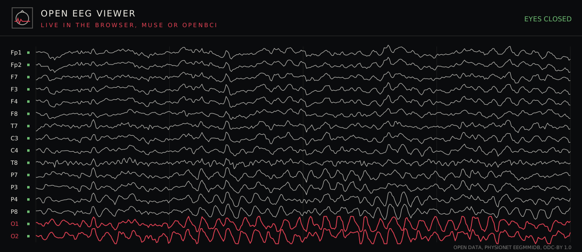
This project published an open DIY EEG headset frame that holds 16 dry electrodes at Fp1, Fp2, F3, F4, F7, F8, C3, C4, T7, T8, P3, P4, P7, P8, O1 and O2, each hub engraved with its 10-20 name. Its CAD code places each hub with the 10% and 20% steps of this article on a model of the head. The frame is not printed yet, and its design page lists what is checked and what is open. Both the frame and a Muse can be watched live in the browser with the Open EEG Viewer, which draws each electrode under its 10-20 name and maps the alpha rhythm over the head. Ready made headbands and EEG boards are in the brain kits section.
What electrode placement has to do with consciousness
Placement decides which brain rhythms an electrode can see. The alpha rhythm, near 10 Hz, is strongest over the occipital positions O1 and O2 when the eyes close. Under propofol anaesthesia, alpha power moves from the back of the head to the frontal positions, a pattern known as anteriorization, which Patrick Purdon and colleagues tracked in healthy volunteers as they lost consciousness (PNAS, 2013). A recording that has only frontal electrodes cannot show that shift, and one that has only occipital electrodes cannot show where the alpha went. The States of Consciousness console on this site plays 154 minutes of that open 64 channel propofol EEG, so the shift can be followed in a real recording.
The same placement logic sets the limits of measures of consciousness built on EEG. Melanie Boly and Giulio Tononi’s study of pure presence in meditation under integrated information theory used continuous high-density EEG and reported gamma suppression in posteromedial cortex, a finding that needs many electrodes over the back of the head. A 16 position map covers the main lobes, and a 64 or 256 position map resolves finer regions. For the wider science of what any measurement can and cannot tell about consciousness, the overview of how scientists race to define AI consciousness collects the current tests and their limits.
Limits of the 10-20 system
The system places electrodes by fractions of skull measurements, and the brain region under a given point still differs between people. Studies that need exact regions combine EEG with an MRI of the same head. The four landmarks also take care to find, and an error at one landmark shifts every position on its arc. For home and teaching use, the 10-20 map gives positions that are close enough to compare recordings and to see the main rhythms, the alpha rhythm, sleep spindles and the slow waves of deep sleep.